Figures
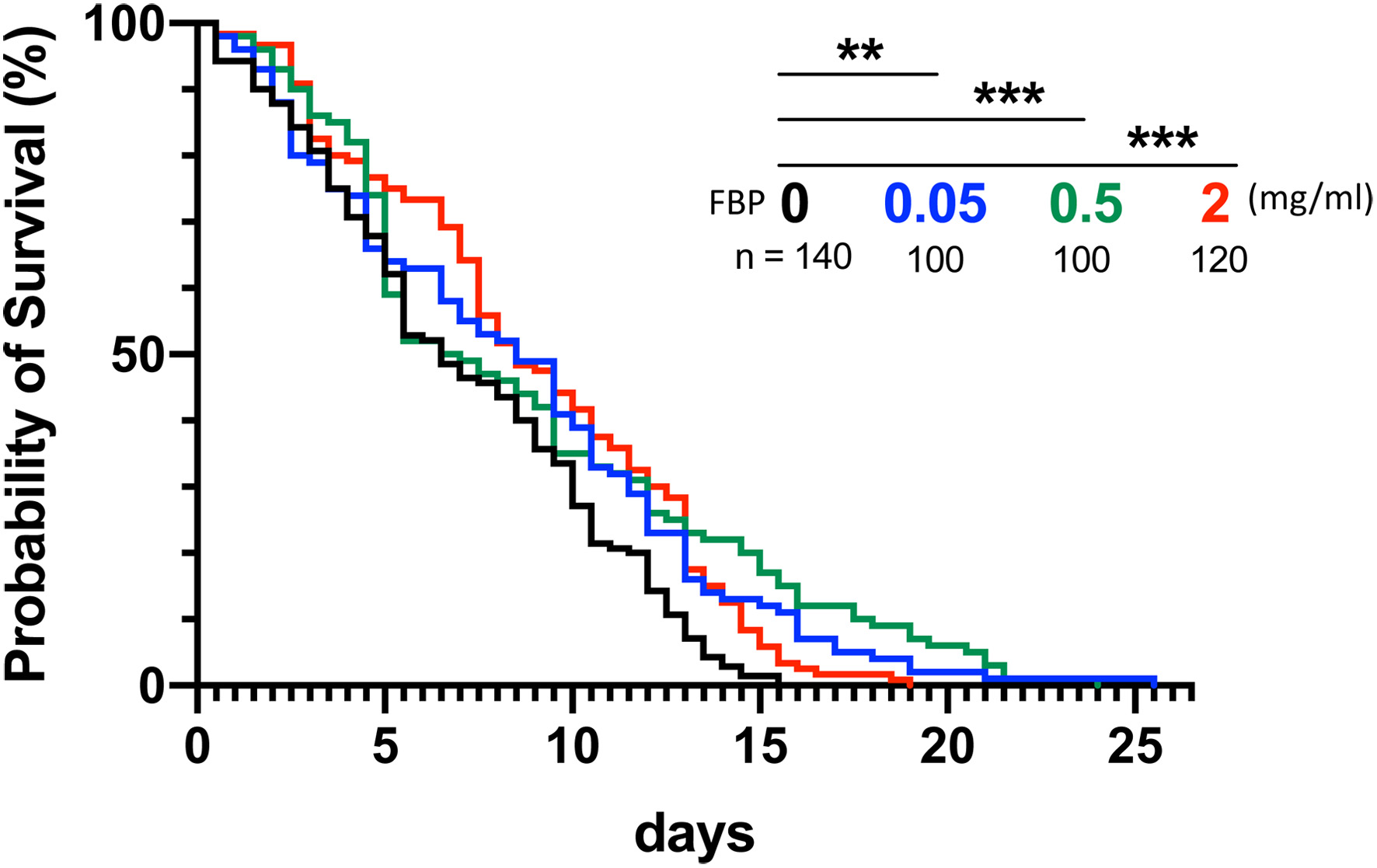
Figure 1. Extension of the lifespan in the Drosophila senescence-accelerated model flies by FBP administration. (A) Lifespan curves in the adult flies fed on the Drosophila instant medium without Fermented Botanical Product (FBP) (0 mg/mL, control, black) or that supplemented with FBP 0.05 mg/mL (blue), 0.5 mg/mL (green), 2 mg/mL (red) of FBP, n ≥ 100 for each. Among twenty young flies collected within 24 hours after eclosion in each vial, dead adults were scored every 12 hour. The results represent the average of five or more than five repeated experiments. Curves were plotted using Kaplan-Meier survival analysis. A log-rank test was performed for adults fed with the control diet and the FBP-containing diet. Note that a significant lifespan extension was observed in every FBP concentration, ns not significant, * p < 0.05, ** p < 0.01, *** p < 0.001, log-rank test.
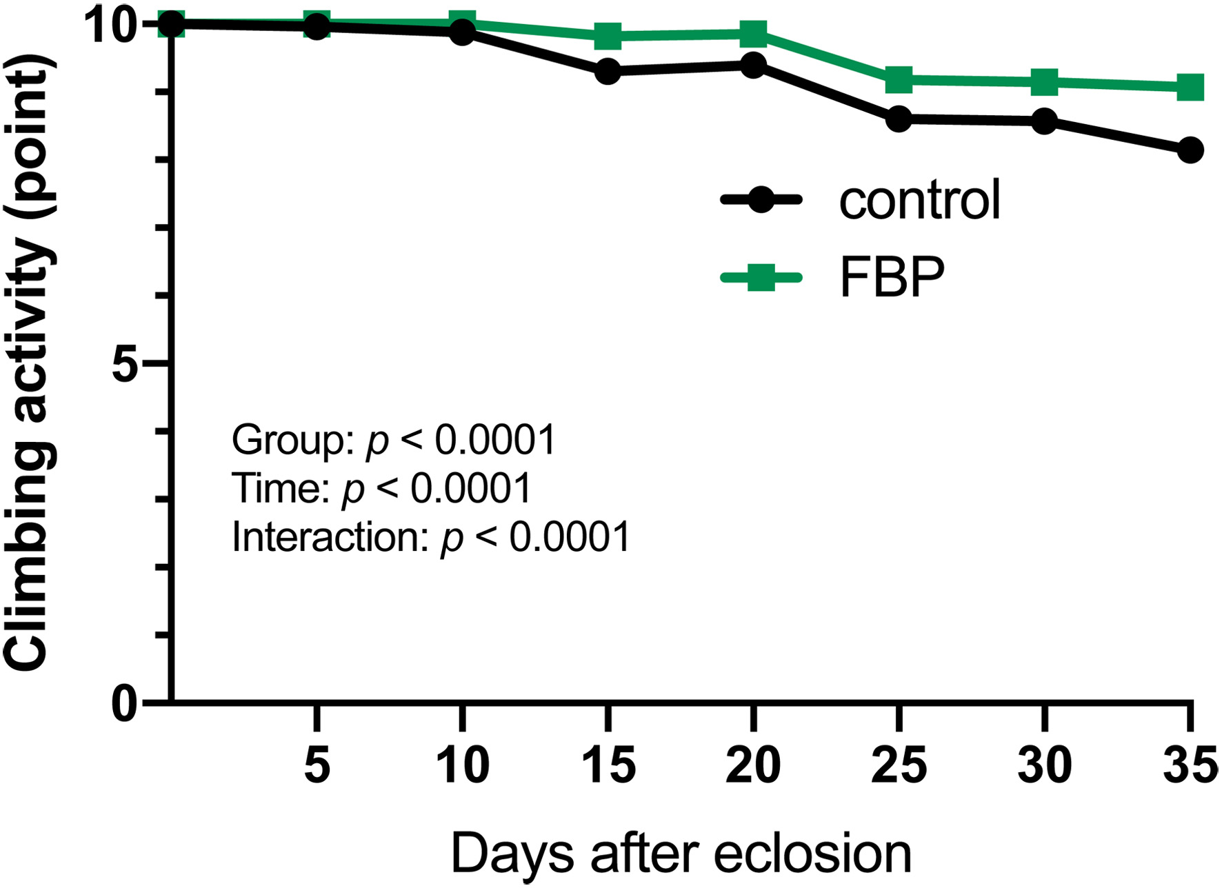
Figure 2. Climbing assay of adults harboring muscle-specific accumulation of oxidative stress by depletion of Sod1 mRNA. Climbing assay to quantify the locomotor activity of adults harboring muscle-specific silencing of mRNAs encoding Superoxiside dismutase 1 (Sod1) (Mef2>Sod1RNAi). Young adults were raised on a diet without FBP (control, black line) or 0.5 mg/mL FBP (green line). Climbing activity was examined every 5 day until 35 days after eclosion (n = 100 from five repeated assays). The points on the y-axis represent the mean climbing scores, which reflect the locomotor activity of the flies. Two-way ANOVA with Tukey’s multiple comparison test. There are significant differences (p < 0.0001) between the treatments for those fed with or without FBP. There are also significant differences in time (p < 0.0001) and treatment-time interactions (p < 0.0001).
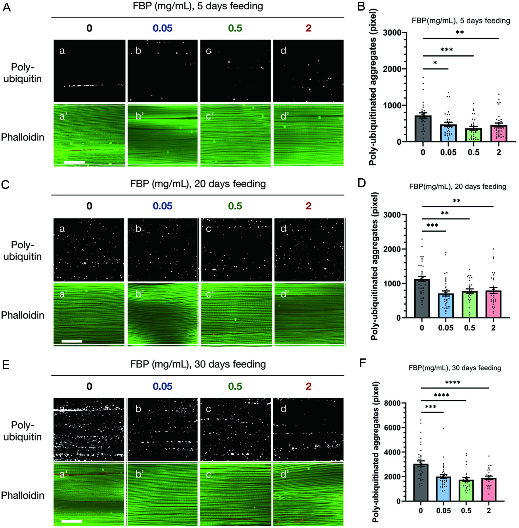
Figure 3. FBP suppresses the accumulation of ubiquitinated protein aggregates in the myofibrils of the indirect flight muscles. (A, C, E) Immunostaining of the indirect flight muscles collected from flies with a muscle-specific depletion of Sod1 (Mef2>Sod1RNAi) using an anti-ubiquitin-conjugated antibody (white) (a-d), and phalloidin staining for F-actin (green) (a′-d′). Young flies, collected within 24 hours after eclosion, were aged for 5 (A, B), 20 (C, D), and 30 (E, F) days on a diet supplemented without FBP (control) (a-a′), or with 0.05 (b-b′), 0.5 (c-c′), and 2 mg/mL FBP (d-d). Scale bar represents 30 μm. (B, D, F) The average pixel total areas of the sum of protein aggregates containing ubiquitinated proteins per single confocal optic fields in control (grey bars), 0.05 mg/mL (blue bars), 0.5 mg/mL (green bars), and 2 mg/mL (red bars) FBP-fed flies are shown on the y-axis. *p < 0.05, **p < 0.01, ***p < 0.001, **** p < 0.0001. One-way ANOVA followed by the Bonferroni post-hoc test was used to assess the differences. The error bars represent the standard error of the mean (SEM).
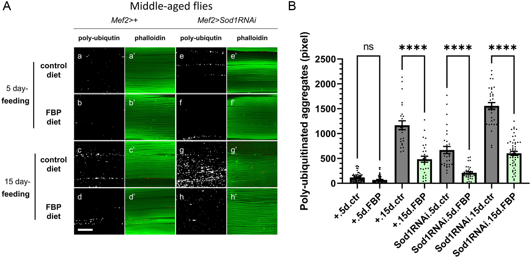
Figure 4. FBP suppresses the accumulation of ubiquitinated protein aggregates in the myofibrils of the indirect flight muscles in middle adult age. (A) Immunostaining of the indirect flight muscles dissected from normal flies (Mef2>+) (Aa-Ad′) or flies with additional oxidative stress accumulation in muscle by the Sod1 depletion (Mef2>Sod1RNAi) (Ae-Ah′) using an anti-ubiquitin-conjugated antibody (white) (Aa-Ah), and phalloidin staining for F-actin (green) (Aa′-Ah′). Young flies, collected within 24 hours after eclosion, were aged on a standard fly food without FBP for 15 days, and then aged on a diet supplemented with or without FBP for 5 (Aa-Ab′, Ae-Af′) or 15 (Ac-Ad′, Ag-Ah′) days. Flies were raised on a diet supplemented without FBP (control) (Aa-Aa′, Ae-Ae, ′ Ac-Ac′, Ag-Ag′), or with 0.5 mg/mL FBP (Ab-Ab′, Af-Af′, Ad-Ad′, Ah-Ah′). Scale bar represents 30 μm. (B) The average pixel total areas of the sum of protein aggregates containing ubiquitinated proteins per single confocal optic field in control (grey bars), 0.5 mg/mL FBP (green bars)-fed flies are shown on the y-axis. One-way ANOVA followed by the Bonferroni post-hoc test was used to assess differences, ns; not significant, ****p < 0.0001. The error bars represent the SEM.
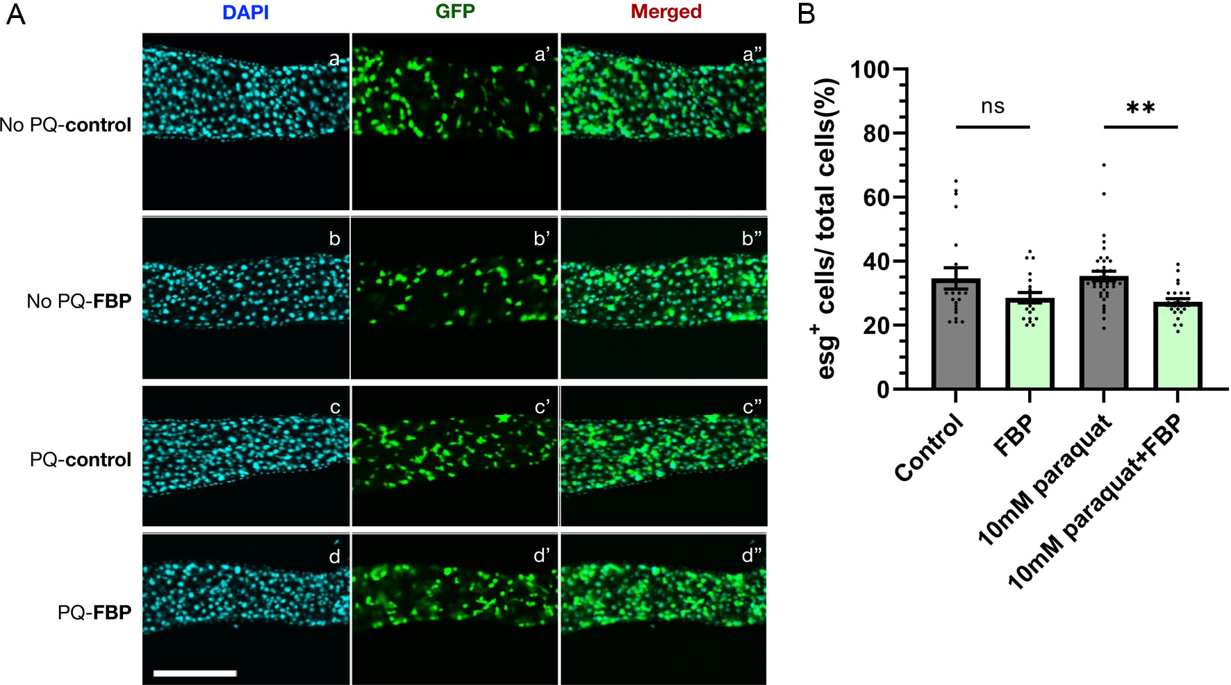
Figure 5. FBP inhibits hyperproliferation of intestine stem cells (ISCs) in intestinal epithelia in aged flies fed with paraquat. (A) Green fluorescence protein (GFP) fluorescence of ISCs expressing GFP in the mid-gut epithelia of esg>GFP flies. Newly eclosed young flies were aged on a standard fly food for 20 days at 28°C, divided into two groups and fed instant medium supplemented with 0.5 mg/mL FBP (Ab-Ab″), or without FBP (Aa-Aa″) for 10 days. For the paraquat-feeding, the 30-day-old flies were incubated in 10 mM paraquat (Ad-Ad″) or without paraquat (Ac-Ac″) for 24 hours. (B) For a quantitative analysis of cell numbers in the adult midgut, the number of 4′,6-diamidino-2-phenylindole (DAPI)-positive (total cells), esg-positive cells (corresponding to intestinal stem cells (ISC)s) in an area of the posterior midgut were counted. The results are presented as the percentage of esg-positive cells among total cells. ISCs are visualized using esg>GFP reporter (green) and DAPI (blue). Scale bar;100 μm. One-way ANOVA followed by the Bonferroni post-hoc test was applied to assess differences; ns not significant, ** p < 0.01. The error bars represent the SEM.


